Figure 6.
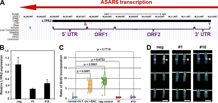
LNA-GapmeR treatment of mouse cells containing the ASAR6 BAC restores normal replication timing. (A) LNA-GapmeR 1 and 10 binding sites along the antisense strand of the L1PA2 within ASAR6. (B) RT-qPCR results from cells transfected with negative control, LNA-GapmeR 1 or LNA-GapmeR 10. A representative experiment, with qPCR reactions performed in triplicate, is shown. Error bars are SD. (C) Replication timing assays on cells treated with negative control, LNA-GapmeR 1, or LNA-GapmeR 10. The ratio of BrdU incorporation in the chromosome 3 with the ASAR6 BAC is compared with the other chromosome 3s in the same cells. The data are visualized using a box plot; the means are shown as horizontal dotted lines, medians as solid lines, and SDs as diagonal dotted lines. The data were analyzed across categories using the ratio of incorporation in multiple cells and the Kruskal–Wallis test, with p-values indicated above the plots. (D) Chromosome 3 images from LNA-GapmeR–transfected cells exposed to BrdU (green) for late replication timing, and then probed for the ASAR6 BAC (top row of chromosomes, purple dot indicated by asterisks). The chromosomes in the bottom two rows in each panel lack the ASAR6 BAC. DNA is labeled with DAPI (blue). Bars, 2 µm.

