
Figure 1. Three proposed pathways for cross-presentation and their regulation by PRRs.
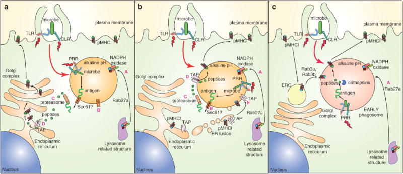
A) The phagosome-to-cytosol pathway. After phagocytosis, antigen is transported out of the phagosome, possibly through the Sec61 translocon. Once in the cytosol, antigen follows the classical MHCI route, consisting of degradation by the proteasome, TAP-mediated entry into the ER and routing to the plasma membrane via the Golgi complex. B) The ERgosome pathway. Phagosomes fuse with (components of) the ER (forming an ERgosome), which brings in TAP and pMHCI displaying self-antigen (shown in blue). Antigens are translocated to the cytosol for processing by cytosolic proteasomes, followed by re-entry into the ERgosome, preferential loading of microbial antigen (shown in green) on MHCI and migration of pMHCI complexes to the plasma membrane. C) The vacuolar pathway. Antigens can be directly processed in the early phagosome by resident cathepsins. Resultant microbial peptides (green) are loaded onto MHCI derived from the plasma membrane (presumably after exchange with self-peptides), which arrive in phagosomes via early recycling endosomes (EREs). After loading of microbial peptides, migration to the plasma membrane takes place by unknown mechanisms. PRRs (TLR and CLR) continue to be engaged (indicated by red lightning bolts) by PAMPs in phagosomes, resulting in signals that may promote these pathways via inducing Rab27a-mediated recruitment of NADPH oxidase from lysosomal structures (A) (which may help preservation of antigenic epitopes by limiting acidification), recruitment of Sec61 to facilitate translocation of antigens into the cytosol (B), enhancement of proteasomal (C) and TAP activity (D) as well as ER recruitment of TAP and MHCI to phagosomes (E).