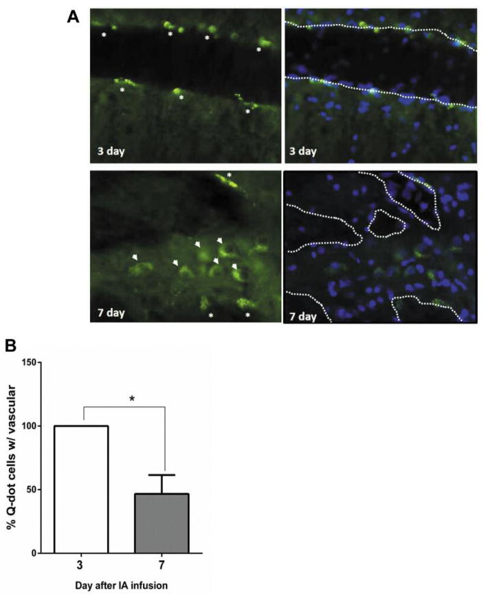
Figure 1.
Cell trafficking of mestatatic breast cancer cells after intracarotid infusion. Athymic rats were intra-carotid infused metastatic MDA-MB-231BR-HER2 breast cancer cells (106) after in vitro labeling with quantum dot (Q-dot) using Qtracker Cell Labeling kit. Brains (n=3 per time point) were harvested from rats at 3 and 7 days after 106 cell infusion. A) Brain vasculature and parenchyma associated Q-dot labeled cancer cells indicated by asterisks and arrowheads, respectively. Brain tissues were counterstained with Hoechst nuclei stain. Putative brain vasculatures were outlined by dashed white line. B) Percentage of vasculature associated Q-dot labeled cells were analyzed from three random fields of view under fluorescent microscopy and represented as mean±SEM. *p<0.05 between 2 groups.

