
Figure 2. TNBC cell migration and proliferation is enhanced by TNBC cell–derived exosomes.
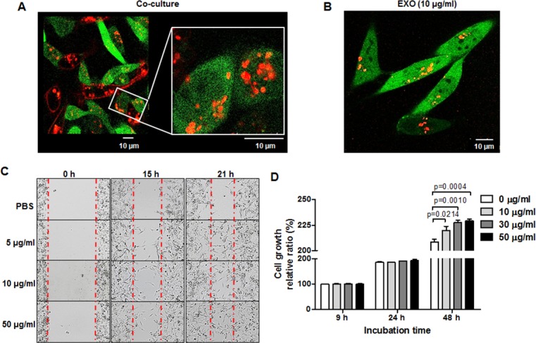
(A) Confocal images of transportation of RFP-tagged exosomes in direct co-culture with MDA-MB-231/CD63-RFP cells and MDA-MB-231/GFP cells for 24 hours. (B) Confocal image of RFP-exosomes (EXO) taken up by MDA-MB-231/GFP cells after administration of RFP-tagged exosomes (10 µg/mL) for 24 hours. (C)Wound-healing assay in MDA-MB-231 cells treated with RFP-tagged exosomes (5–10 µg/mL) or PBS for 15 to 21 hours. (D) Proliferation assay of MDA-MB-231 cells treated with RFP-tagged exosomes (10–50 µg/mL) or PBS for 24 to 48 hours.