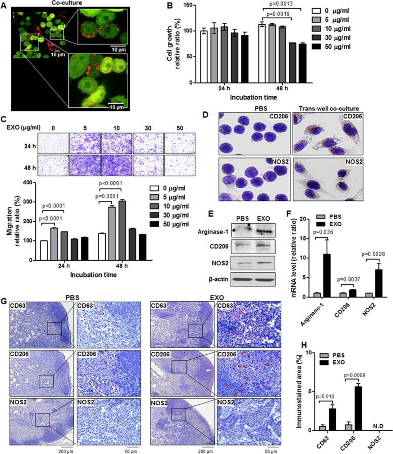
Figure 3. Induction of M1/M2 polarization by TNBC cell–derived exosomes in vitro and in vivo.
(A) Confocal images of RFP-tagged exosome transportation in direct co-culture with MDA-MB-231/CD63-RFP cells and RAW264.7/GFP cells for 24 hours. (B) Proliferation assay in RAW264.7 cells treated with RFP-tagged exosomes (30 or 50 µg/mL) or PBS for 24 to 48 hours. (C) Trans-well migration assay in RAW264.7 cells treated with RFP-tagged exosomes (EXO, 5–10 µg/mL) or PBS for 24 to 48 hours. (D) Immunostaining of CD206 and NOS2 in RAW264.7 cells cultivated with MDA-MB-231/CD63-RFP cells in the trans-well system for 24 hours. (E and F) Western blot and real-time RT-PCR of arginase-1, CD206, and NOS2 in RAW264.7 cells administered RFP-tagged exosomes (10 µg/mL) or PBS for 24 to 48 hours. (G) Immunostaining images of CD63, CD206, and NOS2 in axillary LNs removed from mice at 3 hours after intravenous injection of RFP-tagged exosomes (100 µg) or PBS. (H) Quantitative immunostained area (mean ± S.E.) of CD63, CD206, and NOS2. ND indicates no detection.

