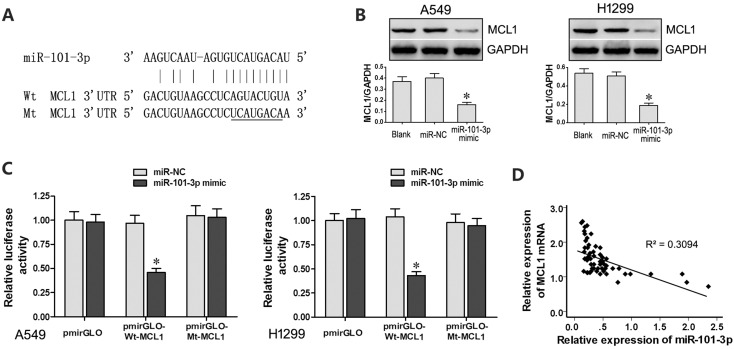
Figure 5. MCL1 is a target of miR-101-3p.
(A) Predicted binding site of miR-101-3p in the 3’-UTR of MCL1 mRNA based on Targetscan analysis. (B) Representative western blots show MCL1 levels in A549 and H1299 cells transfected with miR-101-3p mimics or miR-NC. GADPH was used as internal control. (C) Relative luciferase activity is decreased in cells transfected with Wt-MCL1 (wild type miR-101-3p binding site in 3’UTR) and miR-101-3p mimics than in cells transfected with Mt-MCL1 (mutant miR-101-3p binding site in 3’UTR) and miR-101-3p mimics. This demonstrates that miR-101-3p directly binds to 3’-UTR of MCL1 mRNA. (D) Pearson’s analysis shows correlation between MCL1 and miR-101-3p levels in tissue samples from lung cancer patients sensitive or resistant to cisplatin (n =28/group; Pearson’s co-efficient [R] = - 0.556). Error bars represent mean ± S.D. from triplicate experiments.

