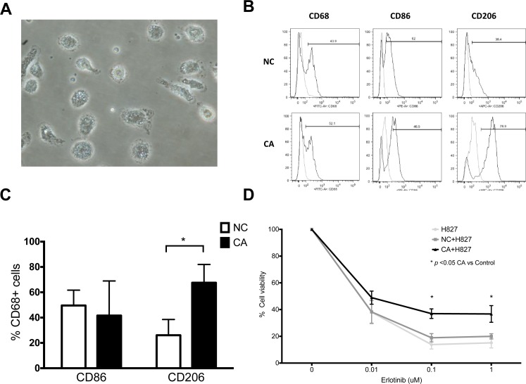
Figure 4. Phenotype and function of bronchoalveolar lavage macrophages.
(A) Representative microscopic image of bronchoalveolar lavage macrophages. 40×. (B) Representative histogram of macrophages from healthy donor (NC) and lung cancer patients (CA). (C) Expression of CD86 and CD206 of bronchoalveolar lavage macrophages. (D) MTT assay cell viability of H827 co-cultured with BALF macrophages from NC and CA. Both cells were treated with designated concentration of erlotinib. n = 7. Figure presented as mean ± SEM. *p < 0.05.

