
Figure 3. Changes in CRF mRNA expression after separation or stressor-exposure in prairie vole mothers.
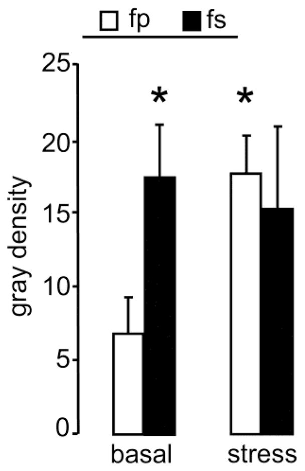
On lactation day 3, prairie vole mothers of each group were either exposed to the forced swim test (see Fig. 3; stress) or left undisturbed (basal). Voles were sacrificed 120 min later, brains were taken, and processed for CRF mRNA in situ hybridization. Expression CRF mRNA in the PVN was increased in stress fp and basal fs mothers compared with basal fp mothers. For further details, see legend to Fig. 1. Data are presented as mean + SEM, n = 5 – 6 per group. * p < 0.05 versus basal fp.