
Figure 2.
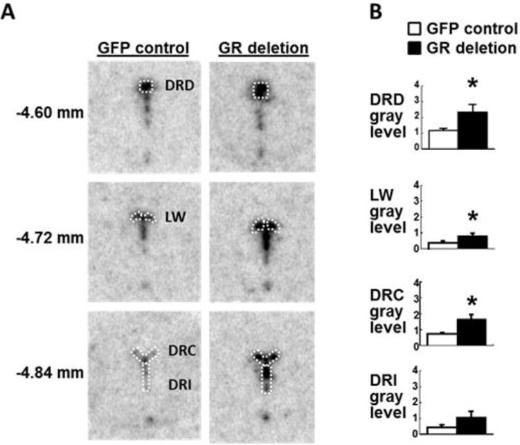
A, Representative examples of tryptophan hydroxylase isoform 2 (tph2) in situ hybridization histochemistry in the DRN of floxed GR mice after DRN injections of AAV2/9 virus transducing green fluorescent protein (GFP control) or Cre recombinase (GR deletion). White outlines show the dorsal DRN (DRD), the lateral wings (LW), the caudal DRN (DRC), and the interfascicular DRN (DRI), as defined by Donner et al. [8]. B, Semi-quantitative analysis of tph2 in situ hybridization histochemistry in GFP control (white bars) and DRN GR deletion groups (black bars). Numbers on the left indicate distance from bregma [29]. Mice were euthanized 3 weeks after DRN injection under basal conditions within 2h of lights-on.
*, P < 0.05 vs. GFP control; n = 5 per group.