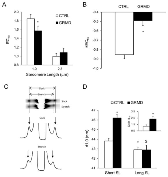
Figure 3. Effect of myopathy on myofilament properties of ENDO permeabilized cardiomyocytes.
Myofilament Ca2+ sensitivity was indexed by EC50 (A) and LDA (B) were evaluated on CTRL (open bars) and GRMD (solid bars) ENDO permeabilized cardiomyocytes at short (1.9μm) and long (2.3μm) SL. (N=4 cells per animal, 4 animals per group). * vs. CTRL; P<0.05. C) Representative X-ray and intensity profile of permeabilized GRMD myocardium as recorded at slack length and after stretch (+20% L0). D) Average myofilament lattice spacing as measured on ENDO myocardium of CTRL (open bars) and GRMD (solid bars) at slack length (Short SL) and after 20%L0 stretch (Long SL). (N=4 animals per group). * vs. CTRL short SL; $ vs. GRMD short SL; P<0.05.

