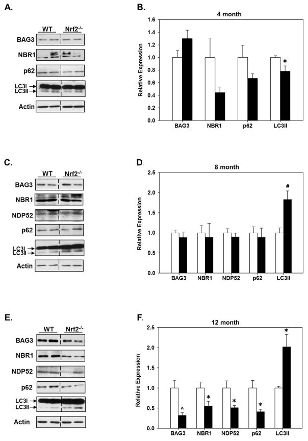
Figure 2. Protein levels of BAG3 and autophagy adaptors are significantly lower in the hippocampus of older Nrf2 (−/−) mice compared to WT mice.
Hippocampal tissue was collected from 4 (A, B), 8 (C, D) and 12 (E, F) month old WT and Nrf2(−/−) mice and immunoblotted for BAG3, NBR1, NDP52, p62 and LC3. Actin served as loading control. Representative immunoblots are shown in (A, C, E) and quantitated data in (B, D, F). At 12 month the levels of BAG3 and all the autophagy adaptors are significantly lower in the Nrf2(−/−) (black bars) compared to the WT mice (open bars), the levels of LC3II are significantly higher. Relative data are expressed as function of WT levels at each age. N=5–6 in each group, mean ± SEM, (*p<0.05, #p < 0.01, ^p < 0.001).

