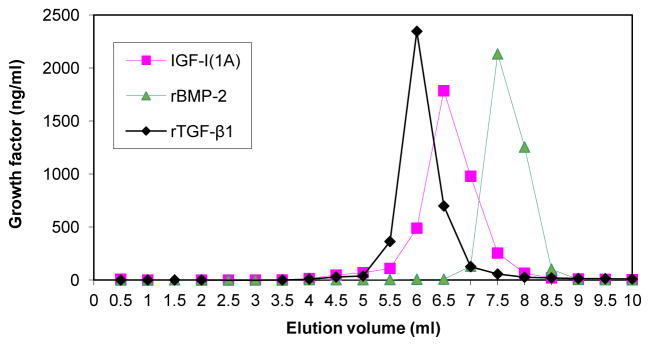
Figure 6.
Comparison of proIGF-IA, BMP-2 and TGF-β1 heparin binding activity. CM from chondrocytes transfected with pAAV-IGF-I1A or PBS containing recombinant BMP-2 (10 ug) or TGF-β1 (8 ug), was analyzed by FPLC using a heparin-sepharose column. CM was applied to the column and the column was washed with 10 ml PBS. The column was eluted with a 10-ml linear gradient of sodium chloride from 0 to 2 M in PBS. The eluate was collected in 0.5 ml fractions. IGF-I, BMP-2 and TGF-β1 in eluate fractions were measured by human IGF-I ELISA, human BMP-2 ELISA and human TGF-β1 ELISA, respectively. Data are expressed as ng/ml of the designated growth factors in each fraction of the eluate.

