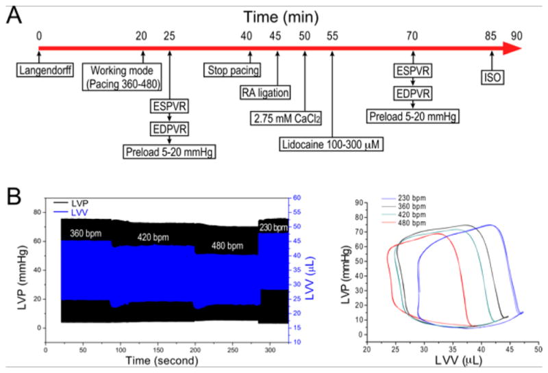
Figure 1. Testing the function of ex vivo mouse working heart at human-like lower heart rate.

A. Protocol steps to test ex vivo mouse working heart function at slower human-like heart rate. B. Representative LV pressure (LVP) and volume (LVV) recordings and P-V loops at 10 mmHg preload and 55 mmHg afterload demonstrate the relationship between the slower heart rate and higher LVP max, lower LVP min, larger end diastolic volume and higher stroke volume.
