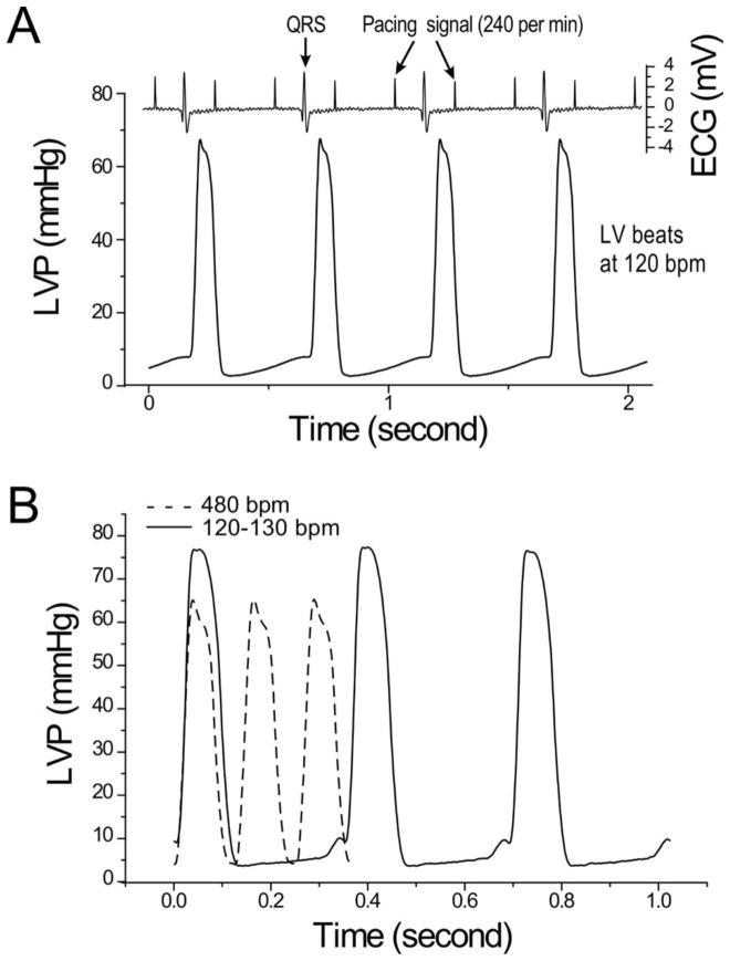
Figure 3. LVP trace of mouse working hearts at a human-like heart rate of 120 bpm.
A. The representative LVP and ECG traces of mouse ex vivo working hearts under 5 mmHg preload and 55 mmHg afterload under 240 per min atrium pacing at and 2.75 mM Ca2+ in 300 μM lidocaine produced 120 bpm ventricular pumping via a 2:1 A-V blockage. B. Comparison between LVP traces of mouse working hearts at 480 bpm at 2.25 mM Ca2+ without lidocaine and 120 bpm at 2.75 mM Ca2+ with 300 μM lidocaine, the LVP waves are similar while the lower heart rate produced an increased peak LVP.

