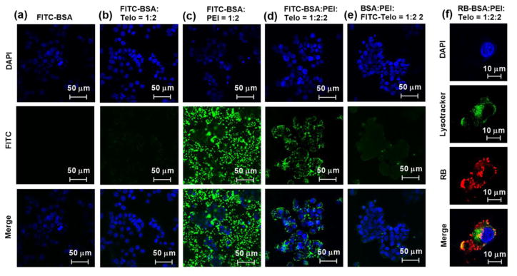
Fig. 7.
Confocal fluorescence microscopy images of (a–e) HT-29 and (f) MDA-231 cells incubated with (a) free FITC-BSA, (b) FITC-BSA:PEG5k(OAOA-L-CHO)4 (1:2, w/w), (c) FITC-BSA:PEI (1:2, w/w), (d) FITC-BSA:PEI:PEG5k(OAOA-L-CHO)4 (1:2:2, w/w), (e) BSA:PEI:FITC-PEG5k(OAOA-L-CHO)4 (1:2:2, w/w), and (f) RB-BSA:PEI:PEG5k(OAOA-L-CHO)4 (1:2:2, w/w) at 37 °C for 3 h. The cell nuclear was stained with DAPI (blue). The lysosome in f was stained with Lysotracker (green).

