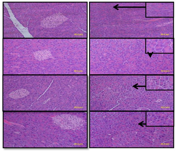
Fig. 2.
Histopathological evaluation of H&E stained pancreatic tissue sections from ethanol-fed ADH− and ADH+ deer mice (bar = 50 μm): A2) & B2) Pancreas of pair-fed control ADH− & ADH+ deer mice, respectively, showing normal histological appearances. C2) Pancreas of ethanol-fed ADH− deer mice showing degenerative changes in the form of individual acinar cell atrophy. D2) Pancreas of ethanol-fed ADH+ deer mice showing only a few acinar cells with degenerative changes. A1, B1, C1 & D1) Pancreas of ethanol-fed and control groups showing relatively well-preserved and intact islets of Langerhans.

