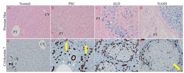
Figure 2. Impact of PSC/IBD on hepatic iron accumulation and its correlation to oval cell proliferation. Panels.
A–D Tissue sections from normal, end-stage PSC/IBD, end-stage ALD and end-stage NASH were evaluated for iron accumulation using Prussian blue staining. Panels E–H. Cytokeratin 7 (CK7) staining of normal, PSC/IBD, ALD and NASH sections (Yellow arrows indicate hepatocyte-like cells that are CK7 positive). Figures are representative of hepatic tissue isolated from at least 4 normal, and 6 PSC/IBD, 6 ALD and 6 NASH patients respectively. All images are 200x magnification (PT=portal triad, CV-central vein).

