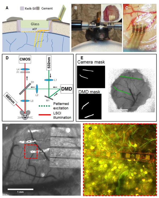
Fig. 1. Nanoelectronic thread enabled multimodal neural platform.
A: Schematic of skull fixation showing the chronic optical access and NET implantation at the surface and cortical depth. Not drawn to scale. B: Photograph of a typical mouse with implanted NET probes and a glass window mounted on top on a customized treadmill for awake recording. Insets: zoom-in view of the glass window in which the arrow denotes an implanted probe. C: photograph of a mouse brain showing that three shanks of NETs implanted intracortically and one on the surface. D: Schematic of the imaging system consist of a laser speckle imaging using 685 nm illumination and a diode lasers (532 nm) coupled with the DMD to provide structured illumination for targeted photothrombosis. E: Example of the image transformation used for DMD pattern projection that allows precise targeting of individual branches of arterioles. F: A representative LSCI near implanted NETs. G: Stacks of two-photon imaging showing little perturbation of the NET on local neuronal(yellow) and vascular (green) networks. Neurons are fluorescently labeled by virus transduction (turbo-RFP) during NET implantation.

