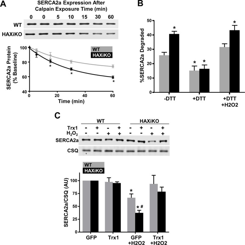
Figure 5. HAX-1 Regulates SERCA2a degradation in SR microsomes and cardiomyocytes in a redox dependent manner.

SR microsomes were isolated from WT and HAXiKO hearts and incubated with purified calpain 1 and 2 mM CaCl2 for indicated times at 37 C. (A) Representative Western blots and quantification of SERCA2a expression. (B) The extent of SERCA2a degradation in SR microsomes after 60 minutes of calpain exposure from WT and HAXiKO hearts is shown under: low reducing conditions (-DTT); high reducing condition (+DTT); and high reducing conditions followed by resuspension in non-reducing buffer and treated with H2O2 (+DTT/H2O2) (n = 3). (C) Cardiomyocytes were isolated from WT and HAXiKO hearts, infected with GFP or thioredoxin 1 (Trx1) adenoviruses, and exposed to H2O2 for 1 hr at 37 C. Representative Western blots of SERCA2a expression levels and corresponding quantification. Calsequestrin was used as a loading control (n = 3). Data are presented as mean ± SEM (P < 0.05: * vs WT GFP/-DTT, # vs WT GFP H2O2).
