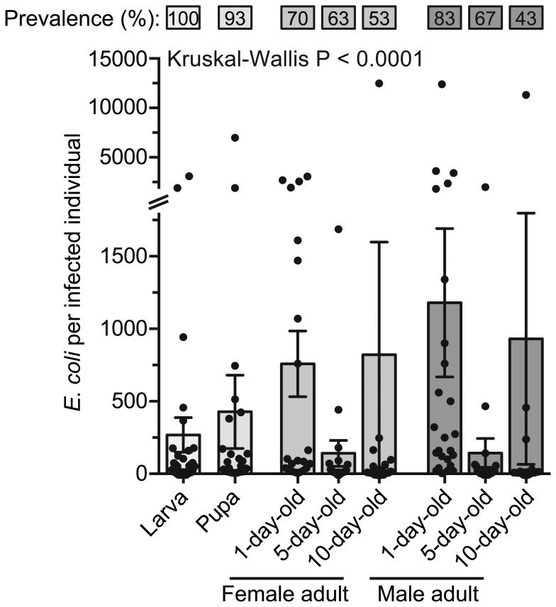
Figure 2.
Prevalence and infection intensity of individuals infected as larvae. Mosquitoes were intrathoracically infected with E. coli and the percentage of mosquitoes infected (top) and the number of E. coli in infected mosquitoes (bottom) was quantified. Column heights mark the mean, whiskers denote the S.E.M, and circles denote the values for individual mosquitoes. Kruskal-Wallis P-value compares infection intensity across stage and age.

