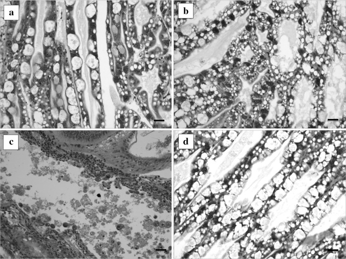
Fig. 1.
Histopathological features of the hepatopancreas of shrimps at 48 h of phage treatment, the shrimp was challenged by AHPND-V. parahaemolyticus 13-028/A3 strain and treated with the phage pVp-1. Negative control (a) and phage control (b) showed the normal appearance of the hepatopancreas. Positive control (c), challenged but not treated, showed the acute sloughing of hepatopancreatic tubular epithelial cells. The phage-treated shrimp d demonstrated the protected morphology of the hepatopancreas. Scale bars 30 μm

