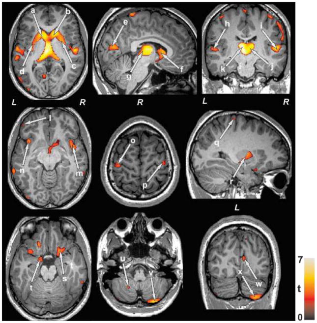
Figure 1.
Brain sites with regional lower gray matter density in SVHD compared to control subjects. Brain regions with reduced gray matter density were observed in the bilateral caudate nuclei (a, b, f), putamen (c, d, r), occipital cortices (e, w), thalamus (g, j, k), temporal gyrus (h, i), prefrontal cortices (l), insular cortices (m, n), precentral gyrus (o, p), post-central gyrus (q), para-hippocampal gyrus (s, t), and cerebellar peduncles (u, v, x), in SVHD over controls. All images are in neurological convention (L = Left; = Right). Color bar indicates t-statistic values, with black/red color shows less significance and yellow color indicates higher significance in gray matter density differences between SVHD and control subjects.

