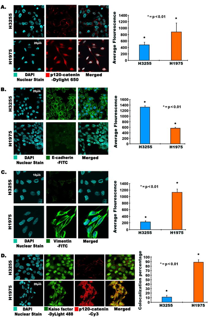
Fig. 2.

(A, B, C, D) Immunofluorescence images showing modulation and changes in morphology of EMT related proteins in TKI-resistant H1975 and H3255 cells. 3×10⁴ cells per well were plated in an 8-well chamber slide, fixed, and probed for (A) p120-catenin, (B) E-cadherin, (C) Vimentin, and (D) colocalization of p120-catenin and Kaiso factor. Colocalization and immunofluorescence was conducted as described earlier[2]. Cells were probed with p120-catenin and Kaiso factor antibodies. DAPI was used as a nuclear stain; DyLight-488 conjugated secondary antibody was used for Kaiso factor, and Cy3 conjugated secondary antibody was used for p120-catenin. Images were captured using an Olympus fv10i confocal microscope. Fluorescence quantification was performed using the Olympus Fluoview image analysis software, and average fluorescence is graphically depicted showing statistically significant results.
