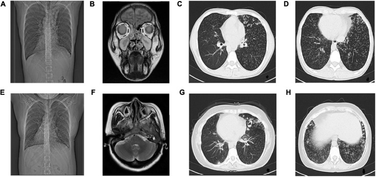
FIGURE 2.

Radiological examination of Proband and Proband’s Sister. The radiological examination of Proband (II-5), (A) CT showed situs inversus. (B) Coronal view of paranasal sinus MRI shows bilateral maxillary sinusitis and ethmoid sinusitis. (C,D) HRCT of chest shows bronchitis and bronchiolitis. Bronchiectasia involves in left lobes. Centrilobular nodules and tree-in-bud sign distribute diffusely in both lobes.The radiological examination of Proband’s sister (II-1), (E) CT showed situs inversus, (F) Transverse view of sinus MRI showed bilateral maxillary sinusitis, (G,H) HRCT of chest showed bronchitis and bronchiolitis, ring-shaped or ductal opacities bronchiectasia involves in both lobes. Secretion obstruction can be seen in some saccular bronchiectasis in the upper left lobes.
