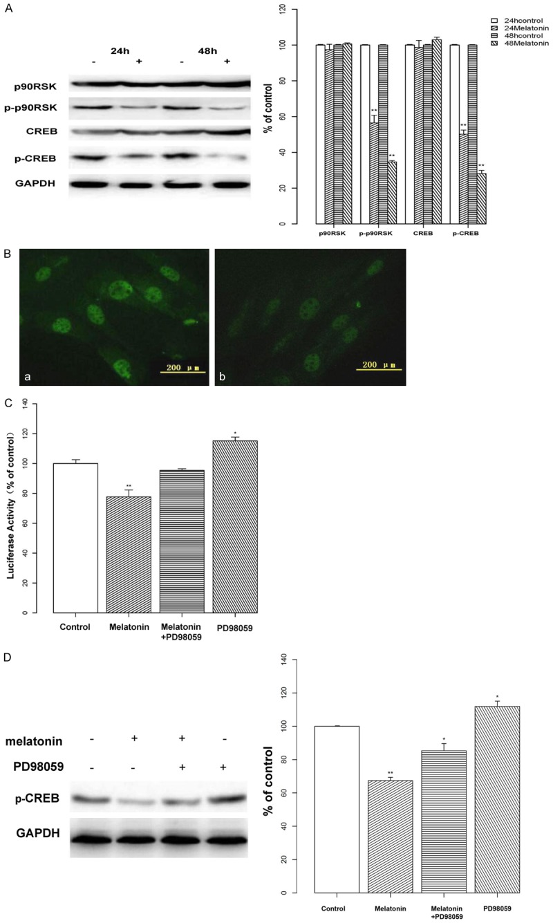
Figure 3.

Effects of melatonin on CREB phosphorylation & transcriptional activity. A: The phosphorylation of p90RSK and CREB were investigated by Western blotting following treatment with melatonin (2 mM; indicated by “+”) or vehicle (indicated by “-”) for 24 or 48 h. Melatonin decreased the phosphorylated expression of p90RSK and CREB in osteoblast cells. Protein levels were analyzed and normalized to the corresponding GAPDH band. Data are displayed as the percent optical density relative to the respective controls. B: Osteoblast cells were treated with vehicle (a) or melatonin (2 mM; b) and stained with a p-CREB antibody. Visualization of the immunofluorescence assay confirmed that treatment with melatonin reduced fluorescent staining, especially nuclear localization. C: Osteoblasts were transiently co-transfected with the luciferase reporter vector pGL3-CRE-luc or with pGL3, and with phRL-TK. After 24 h, cells were treated with melatonin (2 mM) or a MEK inhibitor (PD98059; 50 μM) alone, or in combination for 24 h. CRE-luciferase activity was measured by CRE-luciferase assay. Melatonin treatment reduced the activity of the CRE-luciferase, whereas activity was elevated by PD98059 treatment. D: Osteoblasts were treated with melatonin (2 mM; indicated by “+”) or PD98059 (50 μM; indicated by “+”) alone, or in combination. Protein levels were analyzed by Western blotting and normalized to their respective GAPDH bands. Data are displayed as the percent optical density relative to each protein’s untreated control. Treatment with PD98059 increased the level of CREB phosphorylation. All experiments were performed in triplicate and data are shown as the mean ± S.E.M. *P < 0.05 or **P < 0.01, compared to controls.
