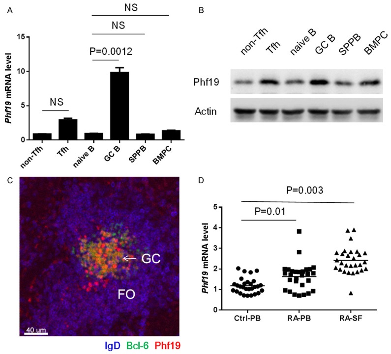
Figure 1.

Phf19 is induced in GC reactions and RA patients. Real-time PCR (A) and Immunoblot (B) analysis of Phf19 expression in mouse splenic naïve B cells, GC B cells, plasma cells, bone marrow plasma cells, follicular helper T cells and non-follicular helper T cells obtained from 5 SRBC immunized C57BL/6 mice. Mouse GAPDH was used as reference control in Real-time PCR analysis and β-Actin was used as loading control for Immunoblot analysis, respectively. (C) Frozen splenic sections were simultaneously stained with anti-Phf19, anti-Bcl-6 and anti-IgD. One typical image was shown here. FO, B cell follicle; GC, germinal center. Original magnification, ×100. Scale bar, 40 um. (D) Real-time PCR analysis of Phf19 expression in peripheral blood (PB; n = 30) and synovial fluid (SF; n = 30) of RA patients and PB (n = 28) of healthy controls. Human GAPDH was used as reference control. Data between two groups were compared and the P value were shown as indicated. NS, not significant. Data are representative of three independent experiments.
