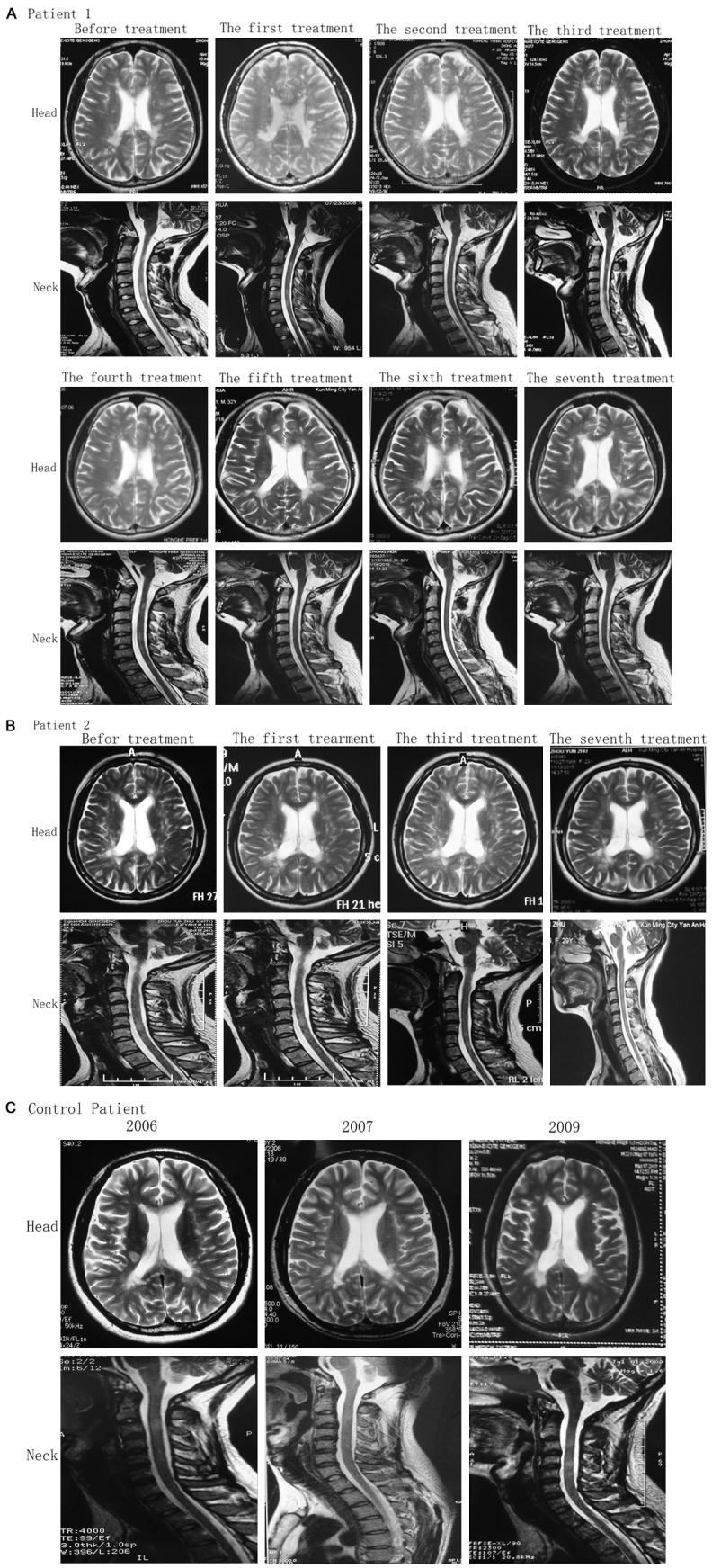
Figure 4.

MRI images of patients in the cerebral transverse plane and sagittal cervical segment at different time points. A. Patient 1, after patient 1 received seven times of treatments, lesions on the right side of the cerebral ventricle became pale and tended to disappear through MRI examination. In addition, the lesions on the left side were remarkably reduced and the ranges of the frontal and parietal lobes and semi-oval area were reduced. The lesion on the cervical spinal cord also became lighter. B. Patient 2, MRI results revealed that the high signal intensity was reduced at the site next to the left ventricle and basal ganglia. C. Patient in the control group, high signal intensity was visible in the white matter lateral to the bilateral ventricles and semi-oval area. Multiple nodular lesions were observed in the corona radiata and the site next to the lateral ventricles.
