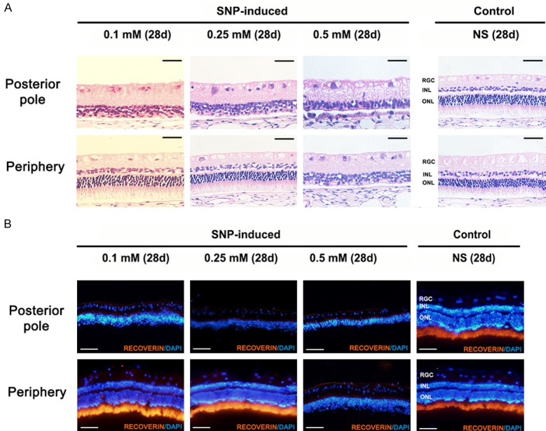
Figure 3.

Hematoxylin-eosin staining and immunofluorescence staining of the retina in New Zealand White rabbits. A. Hematoxylin-eosin staining of retinas at posterior pole and periphery day 28 post-injection. The outer nuclear layer and outer plexiform layer were obviously damaged at posterior pole of retina in three SNP groups and at periphery retina in 0.5 mM group while no obvious damage was found at periphery retina in 0.1 mM, 0.25 mM group and control group. Scale bar: 50 µm. B. Immunofluorescence staining of retinas with photoreceptor marker recoverin at posterior pole and periphery day 28 post-injection. The expression of photoreceptor cell marker recoverin was not observed at the posterior pole of retina in SNP groups and periphery retina in 0.5 mM group but prominently expressed in control group and periphery retina of 0.1 mM and 0.25 mM groups. Scale bar: 100 µm.
