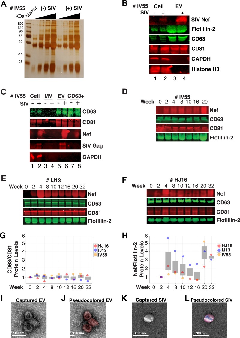
FIG 6 .
Nef is a constituent of CD63+ EVs in vivo. (A) Silver stain analysis of total EVs isolated from macaque IV55 pre- and postinfection with SIV. EVs were diluted to equivalent concentrations (1 × 10⁹, 2 × 10⁹, 4 × 10⁹, and 8 × 10⁹ particles/ml), and contents were run for silver stain analysis. (B) Nef is detected in the EV fraction from SIV-infected macaques. Cell pellets and EV fractions from animal IV55 pre- and postinfection with SIV were assayed for the presence of Nef. Cell pellets were run with equivalent total protein input amounts, as determined by GAPDH and histone H3 levels. EV pellets were run with equivalent total numbers of EV particles, as determined by NTA. The EV markers flotillin 2, CD63, and CD81 were used to identify the presence of EVs. The SIVmac239 Nef antibody was used to track Nef. (C) Nef is present in CD63+ affinity-purified EVs. Cell pellet, MV, total EV, and CD63/CD81 affinity-purified fractions were assayed for the presence of Nef and other protein components. (D) Nef levels in CD63+ EVs throughout the course of infection of macaque IV55. EVs were CD63+ affinity purified at various time points pre- and postinfection with SIV. (E) Same as panel D but for IJ13. (F) Same as panel D but for HJ16. (G) Quantitation of the CD63/CD81 levels of the three animals throughout infection. (H) Quantification of the Nef/flotillin 2 levels of the three animals throughout infection. (I) Representative electron micrograph of CD63+ EVs from animal IV55. Scale bar = 100 nm. (J) CD63+ EVs imaged in panel I pseudocolored red to improve contrast. Scale bar = 100 nm. (K) Representative electron micrograph of SIV particle from animal IV55. Scale bar = 200 nm. (L) SIV particle imaged in panel J pseudocolored blue and red for contrast. Scale bar = 200 nm. See also Fig. S2.

