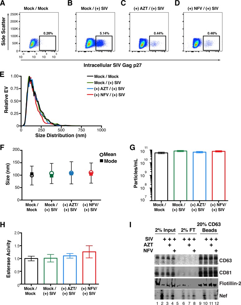
FIG 9 .
Detection of Nef in EVs from SIV-infected primary cells. (A) Primary PBMCs were isolated from rhesus macaques, mock treated, and stained for intracellular SIV Gag (p27) after 5 days. (B) Primary PBMCs were isolated from rhesus macaques, infected with SIVmac239 at an MOI of 10, and stained for intracellular SIV Gag 5 days postinfection. (C) Primary PBMCs were isolated from rhesus macaques and treated with the reverse transcriptase inhibitor AZT (100 nM). Twenty-four hours later, the cells were infected with SIVmac239 at an MOI of 10 and stained for intracellular SIV Gag 5 days postinfection. (D) Primary PBMCs were isolated from rhesus macaques and treated with the viral protease inhibitor NFV (100 nM). Twenty-four hours later, the cells were infected with SIVmac239 at an MOI of 10 and stained for intracellular SIV Gag 5 days postinfection. (E) Size distribution analysis of EVs isolated from the supernatant of simian macaque primary PBMCs infected with SIVmac239 (see Materials and Methods). Videos of EV populations were taken to determine size distributions (11 measurements per group with a total of three biological replicates). The peak size was arbitrarily set to 1 for each group. (F) Mean and mode sizes of EVs from the PBMCs treated for panel A. n = 3 per group. (G) Total EV concentration (particles per milliliter) in supernatant of PBMCs treated for panel A. n = 3 per group. (H) Esterase activity of EVs isolated from the PBMCs treated for panel A. All values are standardized to the Mock/Mock group. n = 3 per group. (I) SIV Nef is present in CD63+ affinity-purified EVs taken from infected PBMCs. Total EVs from panel A were added to CD63 antibody-coated beads and assayed for the presence of Nef. Input, total EV population; flowthrough (FT), unbound fraction; CD63 Beads, contents bound to CD63 beads.

