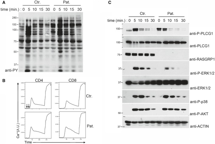
Figure 2. Defective ERK1/2 phosphorylation but normal tyrosine phosphorylation signals and calcium flux in activated RASGRP1‐deficient T cells.

- Immunoblot showing the global tyrosine phosphorylation in T‐cell blasts from a control donor (Ctr.) and P1.1 (Pat.) stimulated with anti‐CD3 antibodies for 0, 5, 10, 15, and 30 min. One representative of two independent experiments from different blood samples.
- Flow cytometry analyses of Ca2+‐flux in T‐cell blasts of a control donor and P1.1 loaded with the Ca2+‐sensitive fluorescent dye Indo‐1. Cells were then stimulated with anti‐CD3 antibody (first arrow) crosslinked with rabbit anti‐mouse antibody (second arrow) and then incubated with ionomycin. Intracellular Ca2+ levels are expressed in arbitrary units (A.U).
- Immunoblots showing phosphorylation of PLCG1 (P‐PLCG1), ERK (P‐ERK 1/2), p38 (P‐p38), and AKT (P‐AKT) in T‐cell blasts from a control donor and P1.1 stimulated with anti‐CD3 antibody for 0, 5, 10, 15, and 30 min. Total ERK 1/2, RASGRP1, and actin (as loading control) are also shown. One representative of three independent experiments from different blood samples.
Source data are available online for this figure.
