Figure 2.

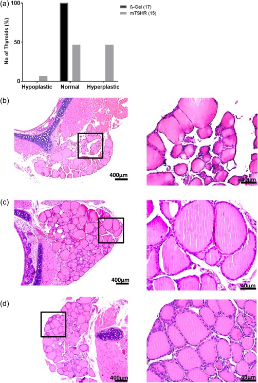
Thyroid histology of mouse thyroid stimulating hormone receptor (TSHR) A‐subunit‐immunized mice. (a) Thyroid slices of the animals were haematoxylin and eosin (H&E)‐stained, and scored for thyroid status as described in the Methods and Results. Histologically scores of β‐Gal and mouse TSHR‐immunized mice were performed blindly by a reader unaware of the immunization scheme. A number of hyper‐, hypo‐ and euthyroid individual mice are shown. All control β‐Gal mice were scored as euthyroid. One mouse TSHR A‐subunit animal was scored as hypothyroid, seven of 15 immune animals as euthyroid and the last seven as hyperthyroid. (b) Thyroid histology of mouse TSHR A‐subunit‐immunized mouse which showed signs of hypothyroidism with characteristically thin follicular epithelium. (c) Example of thyroid of control β‐Gal scored as euthyroid. (d) Thyroid of mouse TSHR A‐subunit‐immunized mouse which demonstrates signs of hyperthyroidism‐like empty follicles and thicker thyrocytes (magnification ×100 and ×400). [Colour figure can be viewed at wileyonlinelibrary.com]
