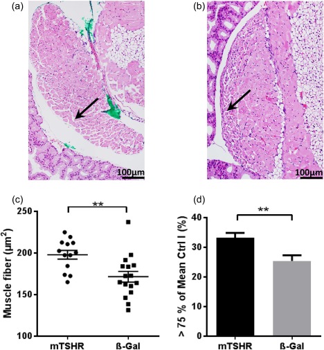
Figure 5.

Muscle changes in the orbital tissue of mouse thyroid stimulating hormone receptor (TSHR) A‐subunit‐immunized mice. Slices of the middle orbital area of one orbit were haematoxylin and eosin (H&E)‐stained to analyse the morphology of muscle. Rectus inferior muscle and obliquus inferior muscle of every individual mouse were analysed quantitatively with Image J to analyse muscle changes. (a,b) representative muscle from mouse TSHR A‐subunit‐immunized animal (magnification ×200). The green colour in A was used as a marker for orientation after preparation of the section. Arrow indicates larger muscle fibres at the edge. (b) An example of a muscle of β‐Gal mouse with an arrow marking smaller muscle fibres at the edge. (c) Further analyses showed that the individual muscle fibres in each muscle are increased in animals undergoing experimental Graves' orbitopathy (GO) (**P ≤ 0·01), but orbital muscles in both groups (mouse TSHR A‐subunit and control β‐Gal) have the same total size (data not shown). (d) After determining the 75th percentile of all control β‐Gal mice muscle fibres (218 µm2) of rectus inferior muscle and obliquus inferior muscle, animals undergoing experimental GO have a statistically significant increased number of larger muscle fibres (**P ≤ 0·01). [Colour figure can be viewed at wileyonlinelibrary.com]
