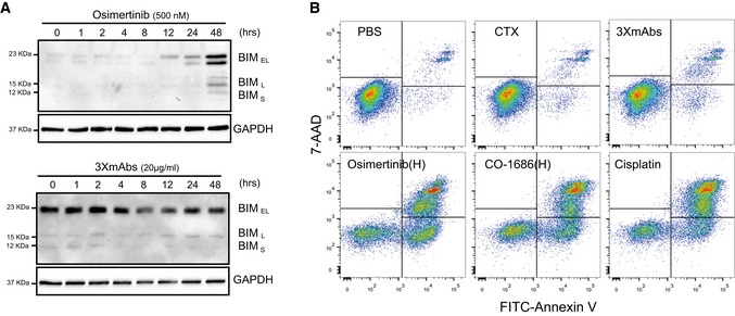
Figure EV2. Unlike antibodies, kinase inhibitors induce apoptosis of mutant EGFR expressing cells.

- PC9‐ER cells were treated with osimertinib (500 nM; upper panel) or with 3×mAbs (20 μg/ml; lower panel) for the indicated time intervals. Cleared cell extracts were subjected to immunoblotting with an anti‐BIM antibody. GAPDH was used as a loading control. The three forms of BIM are indicated.
- PC9ER cells were treated for 48 h with the following agents: saline (PBS), cetuximab (CTX, 20 μg/ml), 3×mAbs (CTX, TRZ, and mAb33, each at 20 μg/ml), osimertinib (0.5 μM), CO‐1686 (0.5 μM), and cisplatin (1 μM). Shown are results of an apoptosis assay performed using an annexin V/7‐AAD kit (BioLegend, Inc). Quantification of the fractions of early and late apoptotic cells are shown in Fig 2C. The experiment was repeated three times.
Source data are available online for this figure.
