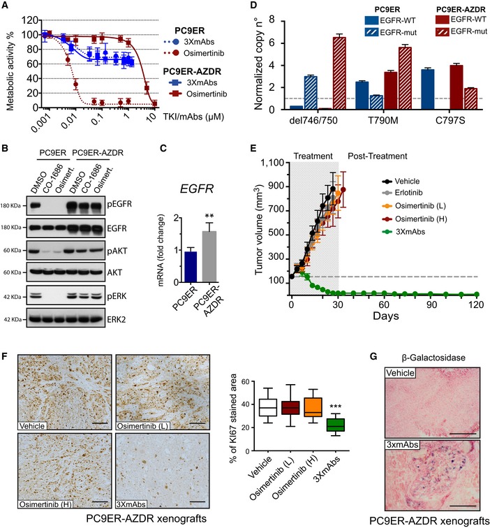
Figure 6. The oligoclonal antibody overcomes the C797S mutation‐mediated resistance to osimertinib.

- Survival assays of PC9ER and derivative PC9ER‐AZDR cells, which were selected in vitro for resistance to osimertinib. Cells were treated for 72 h with increasing concentrations of osimertinib, or with the triple combination of antibodies (3×mAbs; cetuximab, trastuzumab, and mAb33). Data are means ± SD values of three independent experiments.
- Immunoblotting of PC9ER and PC9ER‐AZDR cells treated for 6 h with DMSO (vehicle control), osimertinib (1 μM), or CO‐1686 (1 μM). Whole‐cell extracts were analyzed using electrophoresis and immunoblotting. Blots are representative of two independent experiments.
- Total RNA was isolated from PC9ER and PC9ER‐AZDR cells, complementary DNA was synthesized, and specific EGFR primers were employed. Shown are results of real‐time qPCR analysis after normalization to GAPDH. The assay was repeated three times. Signals represent means ± SD values; **P < 0.05; n = 3; t‐test.
- Copy number variation was determined using digital PCR. Shown are the states of both wild‐type (WT) and EGFR mutations (mut; del746/750, T790M or C797S) in DNA extracted from PC9ER and PC9ER‐AZDR cells. RNASEP was used as normalizer. Error bars indicate the Poisson 95% confidence intervals for each copy number determination.
- PC9ER‐AZDR cells (3 × 10⁶ cells per animal) were subcutaneously grafted in CD1‐nu/nu mice. Thereafter, tumor‐bearing animals were randomized into groups of 9–10 mice, which were later treated once every 3 days with 3×mAbs (0.2 mg/mouse/injection). Alternatively, mice were orally treated with the following TKIs (or vehicle): erlotinib (50 mg/kg/dose) or two doses of osimertinib (1 or 5 mg/kg/dose; L or H, respectively). Tumor growth was monitored once every 3 days. Averages ± SEM values are presented.
- PC9ER‐AZDR cells (3 × 10⁶ cells per animal) were subcutaneously grafted in CD1‐nu/nu mice. Thereafter, tumor‐bearing animals were randomized into groups of 9–10 mice, which were later treated once every 3 days with 3×mAbs (0.2 mg/mouse/injection). Alternatively, mice were orally treated with two doses of osimertinib (1 or 5 mg/kg/dose; L or H, respectively). Shown is immunohistochemical staining of paraffin‐embedded sections for KI67. Scale bars, 100 μm. Also shown are box and whisker plots depicting quantifications of KI67 staining using 6–8 sections/tumor. ***P < 0.001; n = 4; one‐way ANOVA with Tukey's test.
- β‐Galactosidase (β‐Gal) staining of PC9ER‐AZDR tumor xenografts. Tumor‐bearing mice were treated for 20 days with either 3×mAbs (CTX, TRZ, and mAb33; 0.2 mg/mouse/injection, once every 3 days) or saline. Scale bar, 200 μm.
Source data are available online for this figure.
