Fig. 4.
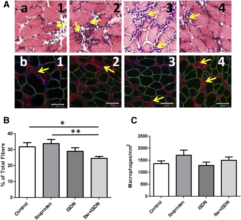
Histologic analysis of necrotic fibers and macrophages in treated muscles. Morphologic studies used H&E to identify and quantitate necrotic fibers, and anti-CD68 antibodies and immunofluorescence to identify and quantitate macrophages. (A) Stained (a) and immunofluorescent (b) images showing regions of muscles on day 3 after LSI under control conditions (1), with ibuprofen (2), with ISDN (3), and with both drugs (4). (B) Necrotic fibers decrease by a small but significant amount at day 3 following injury in mice treated with ibuprofen + ISND, compared with control and ibuprofen alone. **P < 0.01; *P < 0.05. (C) The extent of macrophage infiltration does not differ significantly among groups. Values shown are mean ± S.E.M.

