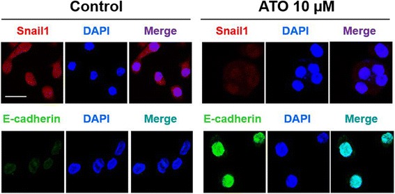
Fig. 2.

Effects of ATO on AGS cells on cellular phenotype and expression of EMT markers. AGS cells were stained with Snail1 stain (red) or E-cadherin stain (green), 4′,6-diamidino-2-phenylindole (DAPI) stain (blue), and after merging respectively. Treatment of ATO 10 μM to AGS cells showed epithelial phenotype with upregulation with E-cadherin and downregulation of Snail1 expression compared with vehicle-treated AGS cells. Note that white bar indicates 20 μm
