Figure 3.

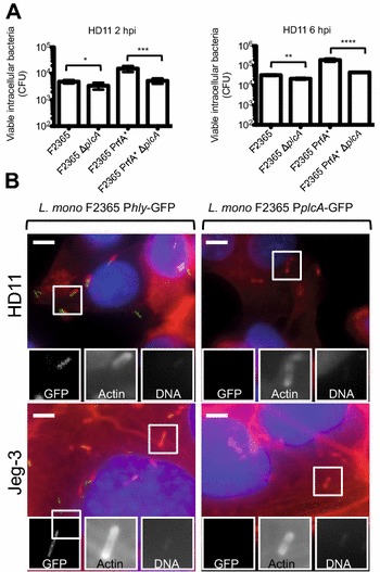
PlcA role and promoter activity in infection of eukaryotic cells. A Number of viable intracellular L. monocytogenes F2365, F2365 ∆plcA, F2365 PrfA* and F2365 PrfA* ∆plcA in HD11 macrophages. CFU numbers were monitored at 2 h and 6 hpi. Indicated are the number of viable intracellular bacteria determined. Three independent experiments with 6 replicates at each experiment were performed. One representative experiment is shown. Means and standard deviation are shown (*, P < 0.05; **, P < 0.01; ***, P < 0.001; ****, P < 0.0001). B Fluorescence microscopy to evaluate the promoter activity of plcA and hly in the L. monocytogenes epidemic strain F2365. HD11, and Jeg-3 cells were cultured in 96 well plates and infected with L. monocytogenes F2365 InlB corrected pAD-PplcA-GFP (right panels) or L. monocytogenes F2365 InlB corrected pAD-Phly-GFP (left panels). Host cells were infected for 6 h and fixed. GFP is shown in green. Actin (red) and nuclei (blue) were labeled with phalloidin conjugated to Alexa 546 and Hoechst, respectively. Bars, 5 µM.
