
Fig. 1.
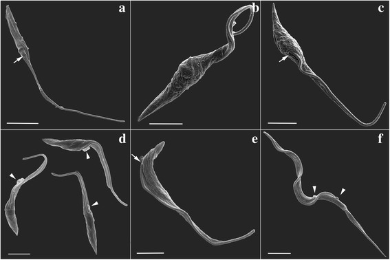
SEM analysis of T. cruzi during metacyclogenesis. a Control epimastigote not subjected to nutritional stress. The flagellum emerges from the anterior portion of the cell body (arrow). b Protozoan subjected to nutritional stress for 2 h. c Adhered cells after 48 h in differentiation medium. The flagellum emerges from the middle of the cell body (arrow). d Cells collected from the supernatant after 72 h in differentiation medium. Arrowheads indicate membrane shedding at the flagellar pocket. e, f Purified trypomastigotes fraction. e Protozoan displaying an epimastigote-like form with the flagellum emerging from the posterior end of the cell body (arrow). f A parasite displaying a typical metacyclic trypomastigote morphology. Arrowheads indicate membrane shedding. Scale-bars: a, 5.0 μm; b-f, 2.5 μm