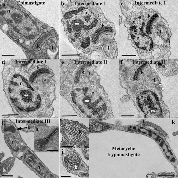
Fig. 5.

Ultrastructural analysis of T. cruzi during metacyclogenesis. Distinct forms are shown: a epimastigote, b-d intermediate I, e, f intermediate II, g, h intermediate III, and k trypomastigote. a-k During differentiation, the kinetoplast is repositioned from the anterior to the posterior end of the cell. g, h The kDNA undergoes topological changes during the late stages of differentiation. i, j Two types of kDNA arrangements are seen in trypomastigotes. Scale-bars: a-g, 0.5 μm; h-j, 0.25 μm; k, 0.5 μm. Abbreviations: f, flagellum; k, kinetoplast; n, nucleus; nu, nucleolus
