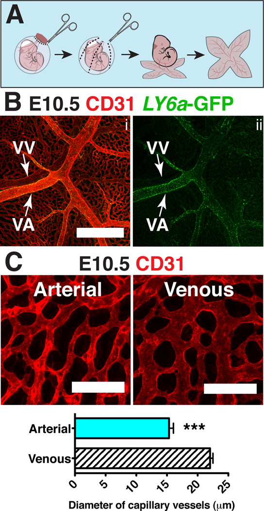
Figure 2. Distinguishing features of arteries and veins in the yolk sac.
(A) Scheme demonstrating removal of the yolk sac prior to imaging to preserve orientation of the vitelline artery and vein. (B) Z-projection of an E10.5 Tg(Ly6a-GFP) yolk sac immunostained for CD31 (i) and GFP (i,ii) . Scale bar = 500µm, VV = vitelline vein and VA = vitelline artery. (C) Z-projection of arterial vascular plexus surrounding the vitelline artery (left) and venous plexus surrounding the vitelline vein (right) at E10.5 in samples immunostained for CD31. Scale bar = 100µm. The diameters of capillary vessels surrounding the vitelline artery and vein were measured using Image J software. Five E10.5 yolk sacs and 60 capillary vessels per yolk sac were measured. The diameter of arterial capillary vessels is 15.3µm ± 0.7µm and the diameter of venous capillary vessels is 22.0µm ± 0.5µm (mean ± SEM). Unpaired 2-tailed Student’s t-test was applied to determine significance. *** indicates that P ≤ 0.001.

