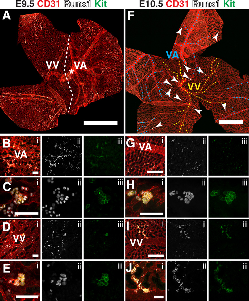
Figure 3. Hematopoietic clusters in the vitelline artery and vein of the yolk sac from E9.5 to E10.5.
(A) Confocal Z-projection of a 22sp yolk sac immunostained for CD31, Runx1 and Kit. Dotted line roughly demarcates the venous and arterial sides of the yolk sac. VA = vitelline artery, VV = vitelline vein. Scale bar = 1mm. (B–E) Confocal images of the vascular plexus near the vitelline artery and vein immunostained for CD31 (i), Runx1 (i,ii) and Kit (i,iii). Scale bars = 50µm. (B) Z-projection of the arterial plexus. (C) Single optical projection of a hematopoietic cluster in the vascular plexus in close proximity to the vitelline artery. (D) Z-projection of the venous plexus. (E) Z-projection of a hematopoietic cluster in the vascular plexus in close proximity to the vitelline vein. (F) Z-projection of an E10.5 yolk sac. Dotted blue lines demarcate the vitelline artery, dotted yellow lines demarcate the vitelline vein, and arrowheads point to CD31+ Runx1+ Kit+ clusters containing 5 or more cells. Scale bar = 1mm. (G–H) Immunostained for CD31 (i), Runx1 (i,ii) and Kit (i,iii). (G) Z-projection of an E10.5 yolk sac near the vitelline artery. Scale bar = 250 µm. (H) Z-projection of a hematopoietic cluster near the vitelline artery. Scale bar = 50µm. (I) Z-projection of the venous vasculature of an E10.5 yolk sac. Scale bar = 250µm. (J) Magnified image of hematopoietic cluster from (I). Scale bar = 50µm.

