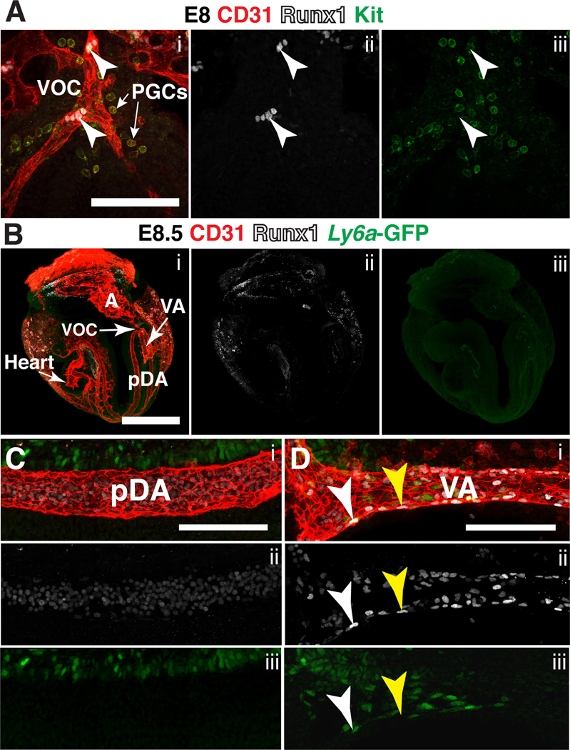
Figure 6. Expression of Runx1 in the major arteries at E8.0 and E8.5.
(A) Confocal Z-projection of the vessel of confluence (VOC) and surrounding primordial germ cells (PGCs) in a late head fold stage embryo (E8.0) immunostained for CD31 (i), Runx1 (i,ii) and Kit (i,iii). White arrowheads point to CD31+ Runx1+ endothelial cells in the VOC. Scale bar = 100µm. (B–D) Immunostaining for CD31 (i), Runx1 (i,ii) and Ly6a-GFP (i,iii). (B) Confocal Z-projection of a 6 sp (E8.5) Tg(Ly6a-GFP) embryo. The top and bottom Z-sections containing the yolk sac were removed to make the vasculature in the embryo proper visible. A = allantois, VA = vitelline artery, pDA = paired dorsal aortae, VOC = vessel of confluence. Scale bar = 500µm. See Movie 2 for animation of Z-stack. (C) Z-projection of one of the two vessels that make up the paired dorsal aortae in an E8.5 Tg(Ly6a-GFP) embryo. Scale bar = 100µm. (D) Z-projection of the vitelline artery; white arrowhead points to a CD31+ Runx1+ Ly6a–GFP+ endothelial cell and yellow arrowhead points to a CD31+ Runx1+ Ly6a–GFP− endothelial cell. Scale bar = 100µm.

