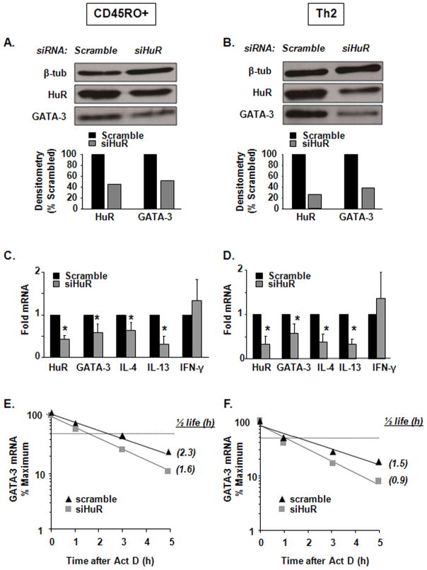
Figure 3. HuR silencing in human CD45RO+ memory T cells and in Th2 cells affects GATA-3 and Th2 cytokine levels.
(A) Western blot (representative of n = 2) of HuR, GATA-3 and β-tubulin protein expression in human CD45RO+ memory T cells and (B) Th2-polarized cells transfected by specific HuR siRNA or a scrambled siRNA control. (C) mRNA steady-state levels of HuR, GATA-3, IL-4, IL-13, IFN-γ measured by real-time PCR in human memory T cells or (D) in human Th2 cells (Mean ± SEM n=3, p<0.05). (E) GATA-3 mRNA decay measured by real-time PCR after treatment with Act D in memory T cells and (E) Th2 cells transfected with HuR siRNA or scrambled control (n = 4; p < 0.05 for half-life). Each experiment (CD45RO+ and Th2 polarization) was performed independently four times (total of eight separate experiments). Western analysis in (A) was performed two times; real time PCR in (C) and (D) three times and actinomycin D in (E) and (F) was performed four times.

