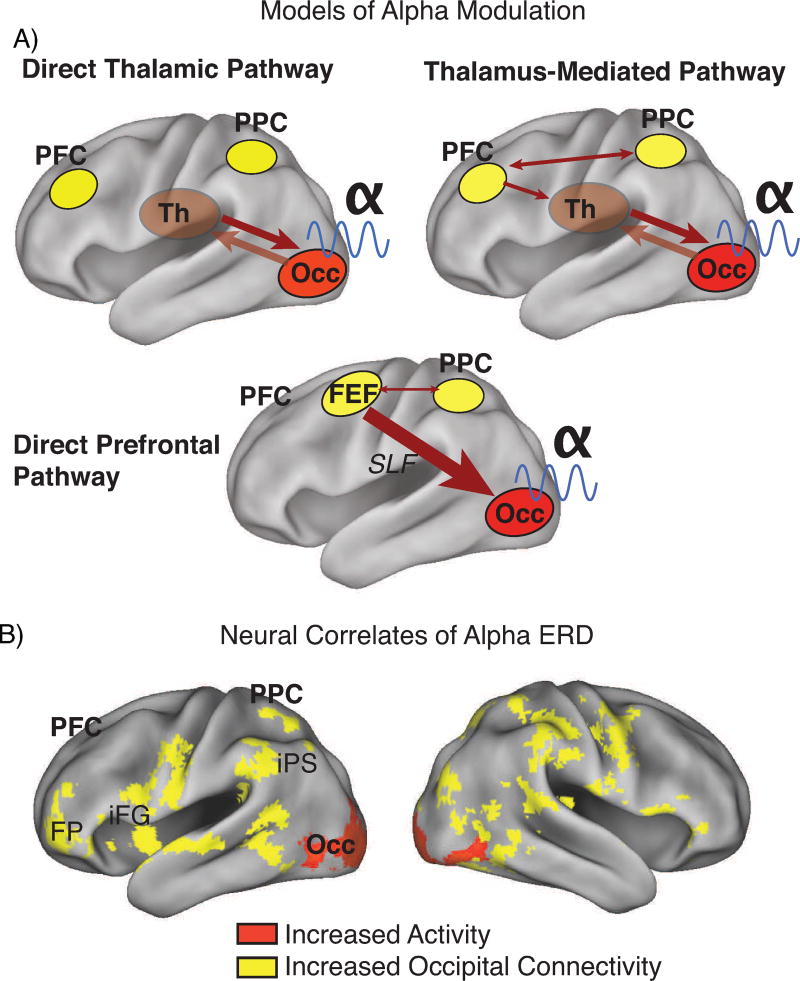
Figure 3. Candidate neural mechanisms of alpha modulation include thalamo-occipital and fronto-parietal interactions.
(A) Modulation of alpha in occipital cortex is likely the result of one of three pathways: bidirectional interactions between occipital cortex and thalamus (Direct Thalamic Pathways), or fronto-parietal interactions exerting top-down influence over occipital activities either via thalamus (Thalamus-Mediated Pathway) or directly (Direct Prefrontal Pathway) via the superior longitudinal fasiculus. (B) Results from a small (n=21) concurrent EEG-fMRI study (121) indicates that alpha ERD in the encoding phase of a spatial working memory trial (c.f., Fig. 2ac) is correlated with both increases in occipital cortex activation and strengthening of functional connectivity between occipital cortex and fronto-parietal regions that include frontal pole, inferior frontal gyrus, post-central sulcus, and, in posterior cortex, intraparietal sulcus and lateral/superior occipital regions. The connectivity also included thalamus (not shown). The data thus support the thalamus-mediated and direct frontal models. Overlays in this image are regression parameters, with threshold at z>2.0, p<0.05 (whole-brain corrected for multiple comparisons using Gaussian random field theory) and mapped to the PALS atlas of human cortex, PFC=prefrontal cortex, PPC=posterior parietal cortex, Th=thalamus, Occ=occipital cortex, iPS=intraparietal sulcus, iFG=inferior frontal gyrus, FP=frontal pole, FEF=frontal eye fields, SLF=superior longitudinal fasiculus.

