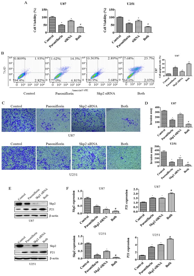
Figure 6.
Effect of Skp2 depletion on glioma cell growth, apoptosis and invasion. (A) Growth of glioma cells treated with PF and transfected with Skp2 siRNA was evaluated with CCK-8. Control, sicontrol; siRNA, Skp2 siRNA; Both, Skp2 siRNA + PF. *P<0.05 vs. control. #P<0.05 vs. either PF treatment or Skp2 siRNA transfection alone. (B) Left panel, apoptosis of U87 glioma cells analyzed by flow cytometry after Skp2 depletion and PF treatment. Right panel, quantitative analysis of results shown in the left panel. (C) Cell invasion was evaluated with the Transwell assay after Skp2 overexpression and PF treatment. (D) Quantitative analysis of results shown in (D). *P<0.05 vs. control. (E) Protein levels of Skp2 and its target P21 as determined by western blotting in glioma cells transfected with Skp2 siRNA and treated with PF. (F) Quantitative analysis of results shown in (E).

