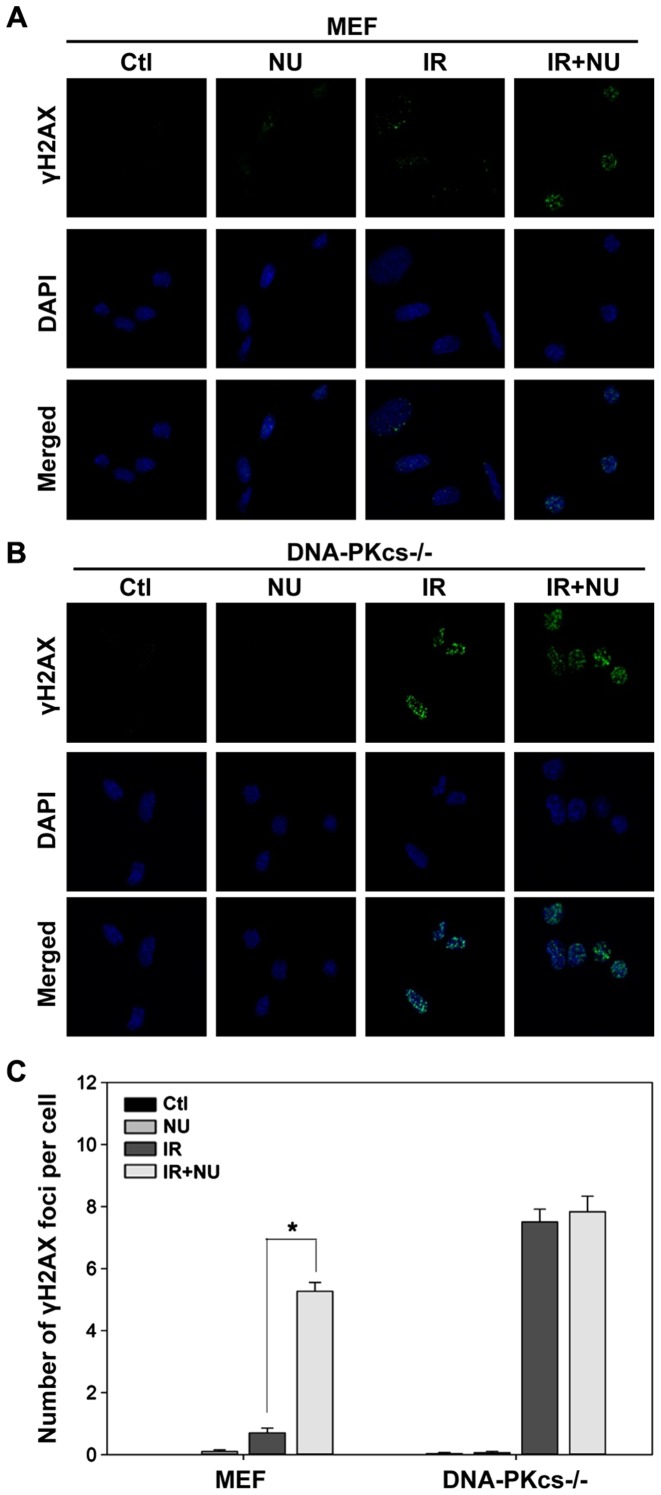
Figure 2.
γH2AX foci formation and its quantification analysis following exposure to ionizing radiation (IR) in the presence or absence of NU7441. (A) WT and (B) DNA-PKcs−/− MEF cells were immunostained with anti-γH2AX antibody 6 h post-IR (5 Gy) in the absence of presence of NU7441 (2 µM). Representative photomicrographs (×1,000 magnification) are shown. (C) Quantification of γH2AX foci. Data are presented as mean ± SEM. *p<0.05, Student's t-test.

