Fig. 2.

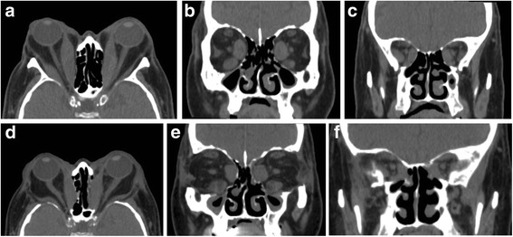
Preoperative and postoperative computed tomography (CT) of the orbit. Preoperative axial (a) and coronal (b and c) CT images showing proptosis, enlargement of extraocular muscles and apical crowding. Postoperative axial (d) and coronal (e and f) CT images showing the reduction in proptosis and relief of apical crowding
