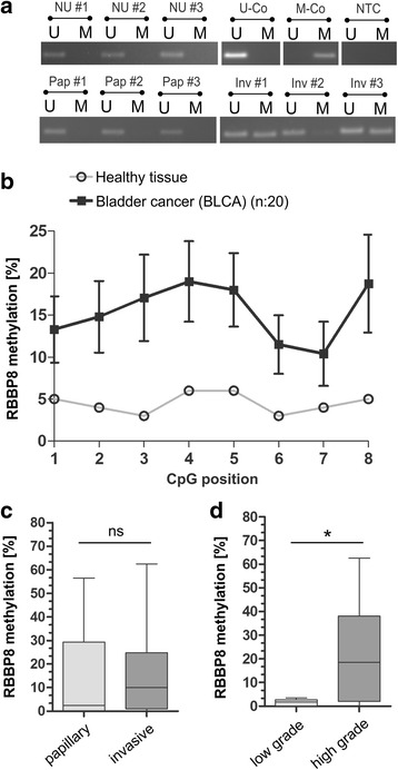
Fig. 4.

Validation of RBBP8 promoter methylation in primary bladder tumors. a Representative MSP analysis shows the RBBP8 promoter methylation status of normal urothelium (NU) and both papillary (Pap) and invasive (Inv) primary bladder cancer tissues. Band labels with U and M represent an unmethylated and methylated DNA locus, respectively bisulfite-converted unmethylated, genomic (U-co) and polymethylated, genomic (M-co) DNA were used as positive controls. NTC: non-template control. b RBBP8 mean methylation values of analyzed CpG sites (1 to 8) of healthy controls and bladder tumors demonstrating tumors-specific hypermethylation. c to d Box plot analysis of RBBP8 methylation in primary bladder tumors is based on mean values of pyrosequenced CpG sites 1–8. c RBBP8 methylation shows no significant differences between the two bladder cancer pathways (papillary and invasive tumors). d Significant enrichment of RBBP8 methylation is demonstrated in high-grade bladder tumors. Horizontal lines—grouped medians. Boxes—25 to 75% quartiles. Vertical lines—range, peak, and minimum; *p < 0.05. Horizontal lines—grouped medians. Boxes—25 to 75% quartiles. Vertical lines—range, peak, and minimum; ns, not significant, *p < 0.05
