Fig. 5.

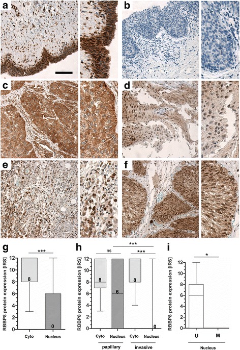
RBBP8 protein loss in nuclei of bladder tumors. Immunohistochemical RBBP8 protein staining of representative tissues are shown. a Strong RBBP8 immunoreactivity was detected in the cytoplasm and in the nuclei of a healthy urothelium, Scale bar: 100 μm. b Negative control of urothelial cell layers. The application of primary antibody was omitted. c Strong RBBP8 immunoreactivity in the cytoplasm of high grade, invasive tumor cells which completely lack nuclear staining. d Moderate cytoplasmatic and heterogeneously nuclear RBBP8 protein staining in invasive tumor cells. e Low RBBP8 protein expression in the cytoplasm of invasive bladder cancer showing strong RBBP8 staining in the nucleus. f Strong nuclear and cytoplasmic RBBP8 staining in non-invasive, papillary tumor cells. g Box plot demonstrating overall significant loss of RBBP8 protein only in the nucleus of bladder tumors. h Box plot graph illustrates the loss of RBBP8 protein within the nuclei of high-grade invasive bladder tumors. i Box plot shows a significant RBBP8 protein loss in tumors harboring RBBP8 promoter methylation. U, unmethylated; M, methylated. Horizontal lines — grouped medians. Boxes — 25 to 75% quartiles. Vertical lines — range, peak, and minimum; ns, not significant, *p < 0.05, ***p < 0.001
